Bicipital Groove X Ray View

Xray View Of Biceps Groove Download Scientific Diagram

Tangential Projection Intertubercular Bicipital Groove

Tangential Projection Intertubercular Bicipital Groove

Arm Humerus X Ray View

Bicipital Groove Radiography Photo Wikiradiography
Joints Radiology Key
Shoulder tangential intertubercular bicipital groove purpose and structures shown.
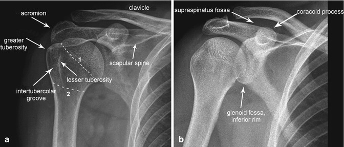
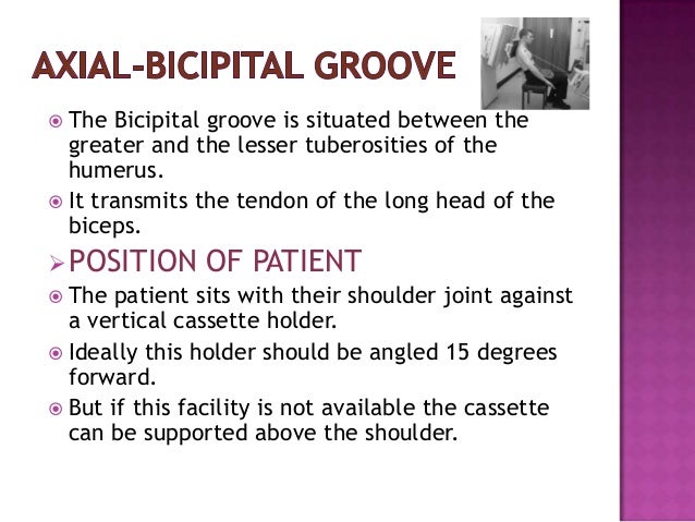
Bicipital groove x ray view. The hand is rotated 45 degrees laterally from prone to bring the bicipital groove in profile centering o the of the beam central ray is directed cranially along the long axis of humerus. The bicipital groove is an osseous groove formed in the humeral head by the medial and lateral tuberosities. Intertubercular groove muscles duration. The stryker notch view is a specialized projection of the shoulder aimed at assessing the posterior humerus.
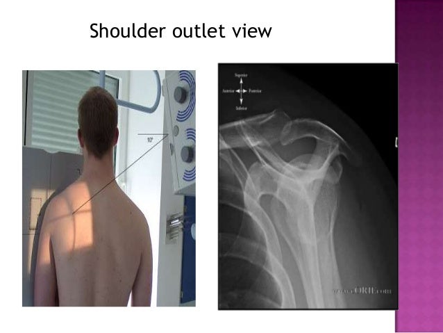
The purpose of this paper was to find a standard radiographic view for measurements of the intertubercular groove of the human humeral head. Indications the stryker notch view can be used post anterior glenohumeral dislocation assessing for hill sachs lesions 1. O centered to the anterior part of the head of humerus. The effect of the direction of the x ray beam on the image of the groove was studied in cadaver specimens.
A standardized optimal x ray beam direction was subsequently used in a clinical study on 30 patients. The shoulder series is fundamentally composed of two orthogonal views of the glenohumeral joint including the entire scapula. It serves to retain the long head of the biceps brachii. The extension of the shoulder series depends on the radiography department protocols and the clinical indications for imaging.
We made special tangential views on 40 such patients and in no instance was a normal groove depicted. Coracoid process humeral head greater lesser tuberosity bicipital groove duration. A presumptive diagnosis of bicipital tenosynovitis is made by taking a thorough history performing a comprehensive physical exam and by reviewing x rays. In this project we investigate the relationship between the 3d shape of the bicipital groove and the incidence of pathology of the long biceps tendon.
X rays may reveal mineralization of the biceps tendon mineralization within the biceps groove or sclerosis hardening of the bone seen as increased whiteness on x rays below the biceps. Bicipital groove x ray see more descriptions. This tool allows you to search snomed ct and is designed for educational use only. Shoulder radiographs are performed for a variety of indications including.
This view should demonstrate the bones and soft tissue of the shoulder specifically the intertubercular groove free of superimposition of the shoulder. Pathologic changes in the bicipital groove region. Bicipital groove x ray bicipital groove x ray procedure hide descriptions.

Anatomically Labelled Ap Shoulder X Ray Medical Knowledge

Medical Imaging Technology Radiographic Anatomy Of Shoulder Joint

Tangential Projection Intertubercular Bicipital Groove
Radiographic Positioning Of Humerus And Shoulder
Upper Limbs Radiology Key

Shoulder Radiology Musculoskeletal Key
Radiographic Positioning Of Humerus And Shoulder

Xray Humerus Tangential Intertubercular Groove Youtube

Humerus Series Radiology Reference Article Radiopaedia Org

Medical Imaging Technology Radiographic Anatomy Of Shoulder Joint
Radiographic Positioning Of Humerus And Shoulder

Shoulder External Rotation View Radiology Reference Article

Shoulder Anatomy And Normal Variants
