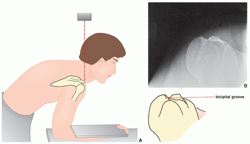
Bicipital Groove X Ray Position

Tangential Projection Intertubercular Bicipital Groove

Tangential Projection Intertubercular Bicipital Groove
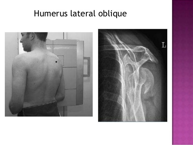
Radiographic Positioning Of Humerus And Shoulder
Radiographic Positioning Of Humerus And Shoulder
Radiographic Positioning Of Humerus And Shoulder

Tangential Projection Intertubercular Bicipital Groove
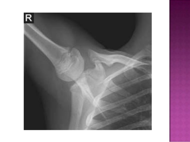
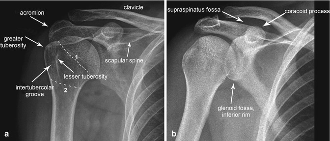
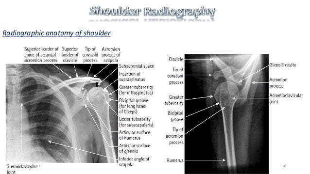
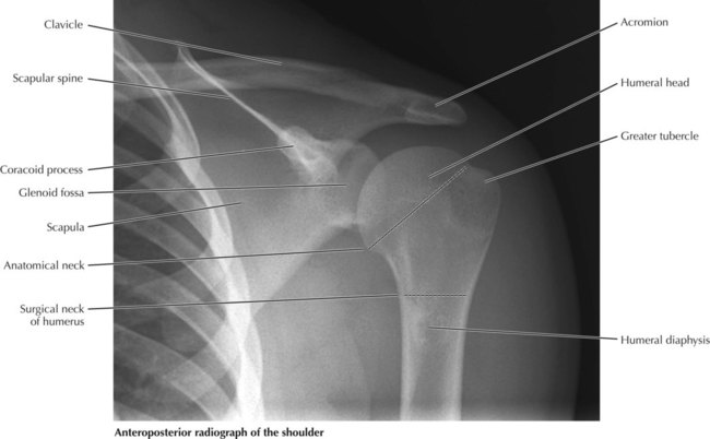
3 1 anteroposterior shoulder radiograph.
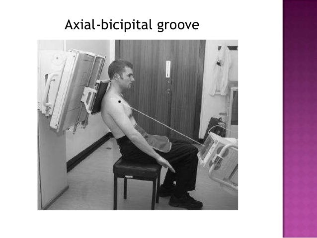
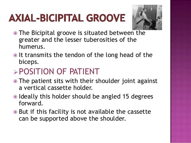
Bicipital groove x ray position. The stryker notch view is a specialized projection of the shoulder aimed at assessing the posterior humerus. The central ray should be 45 degree caudad passing through the scapulohumeral articulation. Indications the stryker notch view can be used post anterior glenohumeral dislocation assessing for hill sachs lesions 1. The purpose of this paper was to find a standard radiographic view for measurements of the intertubercular groove of the human humeral head.
The anterior positioning k wire sets the location for the anterior edge of the plate 2 4 mm lateral to the bicipital groove. Dislocation of the long head of the biceps will inevitably result in rupture of part of the subscapularis tendon. The effect of the direction of the x ray beam on the image of the groove was studied in cadaver specimens. Pathologic changes in the bicipital groove region.
The posterior surface of the injured side should be closest to the the image receptor. Plate too close to the bicipital groove. And no displacement is visible on the x ray then. The hand is rotated 45 degrees laterally from prone to bring the bicipital groove in profile centering o the of the beam central ray is directed cranially along the long axis of humerus.
A standardized optimal x ray beam direction was subsequently used in a clinical study on 30 patients. The proximal positioning k wire determines the proximal edge of the plate 5 8 mm distal to the tip of the greater tuberosity. We made special tangential views on 40 such patients and in no instance was a normal groove depicted. Active assisted forward flexion and.
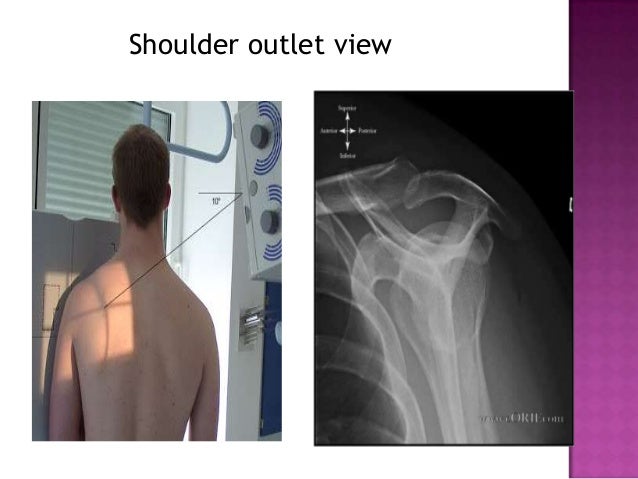
Central x ray should be directed to 2 5 cm inferior to the coracoid process. Anteroposterior shoulder view allows assessment of especially the humeral head lesions and clavicular fractures. Insert k wires through appropriate guiding sleeves. The tendon of the subscapularis muscle attaches both to the lesser tubercle aswell as to the greater tubercle giving support to the long head of the biceps in the bicipital groove.
Ergonomic evaluation of your workstation. While achieving anteroposterior shoulder x ray in neutral position the patient is erect or in supine position. Our cases are limited to patients with a history of previous trauma to the shoulder and ensuing shoulder pain whose routine x ray studies were negative. Conservative treatment of biceps tendonitis is likely to center around reducing the damaging positions in the individual s activities of daily living.
O centered to the anterior part of the head of humerus.
Radiographic Positioning Of Humerus And Shoulder

Shoulder Radiology Musculoskeletal Key

Xray View Of Biceps Groove Download Scientific Diagram

Xray Humerus Tangential Intertubercular Groove Youtube
Joints Radiology Key
Radiographic Positioning Of Humerus And Shoulder

Anatomically Labelled Ap Shoulder X Ray Medical Knowledge
Upper Limb I Shoulder Girdle Radiology Key
Shoulder Radiography Avinesh Shrestha
Upper Limbs Radiology Key

Humerus Series Radiology Reference Article Radiopaedia Org

Shoulder Series Radiology Reference Article Radiopaedia Org
A Representative Post Operative X Ray Examination From The
