Bicipital Groove Mri

Mri Showed Normal Position Of Biceps Tendon In The Bicipital Groove Download Scientific Diagram

Long Head Of Biceps Tendon Dislocation Radiology Reference Article Radiopaedia Org
Long Head Of The Biceps Tear A Mri Arrow Bicipital Groove With No Download Scientific Diagram

Pathology Of The Long Head Of The Biceps Tendon Radsource

Mri Showing The Lhbt Positioned Within The Bicipital Groove Mri Download Scientific Diagram

Proximal Rupture Of Long Head Biceps Brachii Tendon Radiology Case Radiopaedia Org

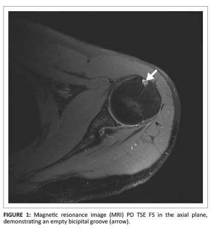
1 axial t1 weighted mr image shows shallow bicipital groove in presence of congenital absence of biceps tendon arrow.
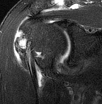
Bicipital groove mri. Classification of the long head of the biceps tendon by arthroscopic findings. High t2 signal intensity noted at the of this humeral head at the biceps groove. The tendon of the subscapularis muscle attaches both to the lesser tubercle aswell as to the greater tubercle giving support to the long head of the biceps in the bicipital groove. High t2 fluid signal intensity is noted around the thickened biceps tendon which is seen within its groove suggestive of tenosynovitis.
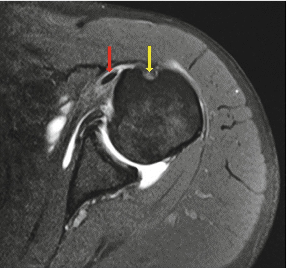
Potential causes of pulley lesions include a congenital rotator interval defect 23 a supratubercular ridge as an osseous protrusion of the lesser tuberosity 24 or a shallow groove 5. Biceps tendon laxity in the bicipital groove may lead to superior extension of degenerative changes in the rotator interval 7 18 20. A type i normal tendon b type ii hourglass shaped hypertrophic tendon with extension of fraying into bicipital groove c type iii partial tear or fraying involving less than 50 of tendon width at the intraarticular region without fraying in the bicipital groove d type iv partial tear involving more than 50 of. The bicipital groove is typically 4 6 mm deep 1.
Biceps tendinitis is a disorder of the tendon around the long head of the biceps muscle. Mri allows preoperative assessment of the lhbt within and distal to the bicipital groove. Bicipital tenosynovitis can be a result of many small tears resulting in inflammation over a period of a number of years or due to an acute injury to the biceps region. Bicipital groove morphology on mri has no correlation to intra articular biceps tendon pathology.
Inflammation of the biceps tendon within the intertubercular bicipital groove is called primary biceps. Thickened with intermediate high t2 signal intensity noted at the supraspinatus tendon suggestive of tendinosis. Dislocation of the long head of the biceps will inevitably result in rupture of part of the subscapularis tendon. The bicipital groove also known as the intertubercular sulcus or sulcus intertubercularis is the indentation between the greater and lesser tuberosities of the humerus that lodges the biceps tendon.
Duplication of the lhbt is a diagnostic dilemma in mri of patients with shoulder pain. It contains the tendon of the long head of the biceps brachii muscle which is ensheathed in a synovial reflection of the. 1 3b orthopaedics university of pennsylvania health system pennsylvania hospital philadelphia pa 19107 usa.
Long Head Of The Biceps Tear A Mri Arrow Bicipital Groove With No Download Scientific Diagram

A Profile View Of A Humeral Head Segmented From A Mri Of The Download Scientific Diagram

Images Of Note 3t Mri Reveals Biceps Tear With Intra Articular Remnant Consult Qd

Mri Showing Dislocation Of The Lhbt Defined As A Complete Loss Of Download Scientific Diagram

Complete Tear Of The Long Head Biceps Tendon Groove Entry Lesion Radiology Case Radiopaedia Org
Pathology Of The Long Head Of The Biceps Tendon Radsource
Complete Rupture Of The Long Head Of The Biceps Tendon And The Distal Biceps Tendon
Axial Magnetic Resonance Imaging Sections Through Bicipital Groove Download Scientific Diagram

Figure 5 From Mri Of The Rotator Interval Of The Shoulder Semantic Scholar
Long Head Of The Biceps Tendon Disease Springerlink

Artifacts And Pitfalls In Shoulder Magnetic Resonance Imaging

Tenosynovitis Of Biceps Tendon Radiology Case Radiopaedia Org
Intertubercular Sulcus Bicipital Groove
