Bicipital Groove Image
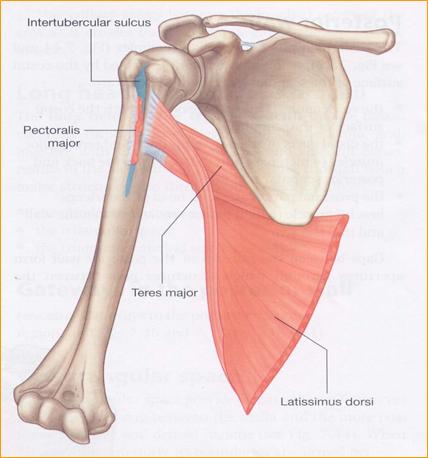
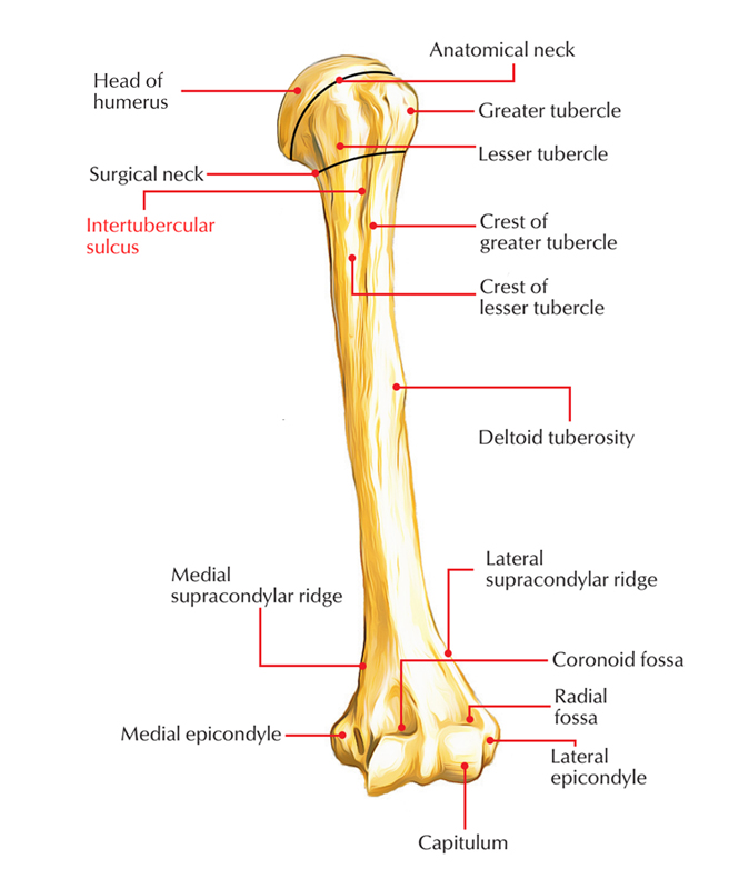
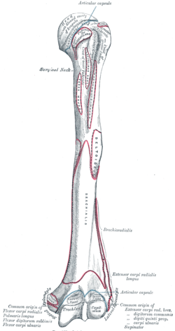
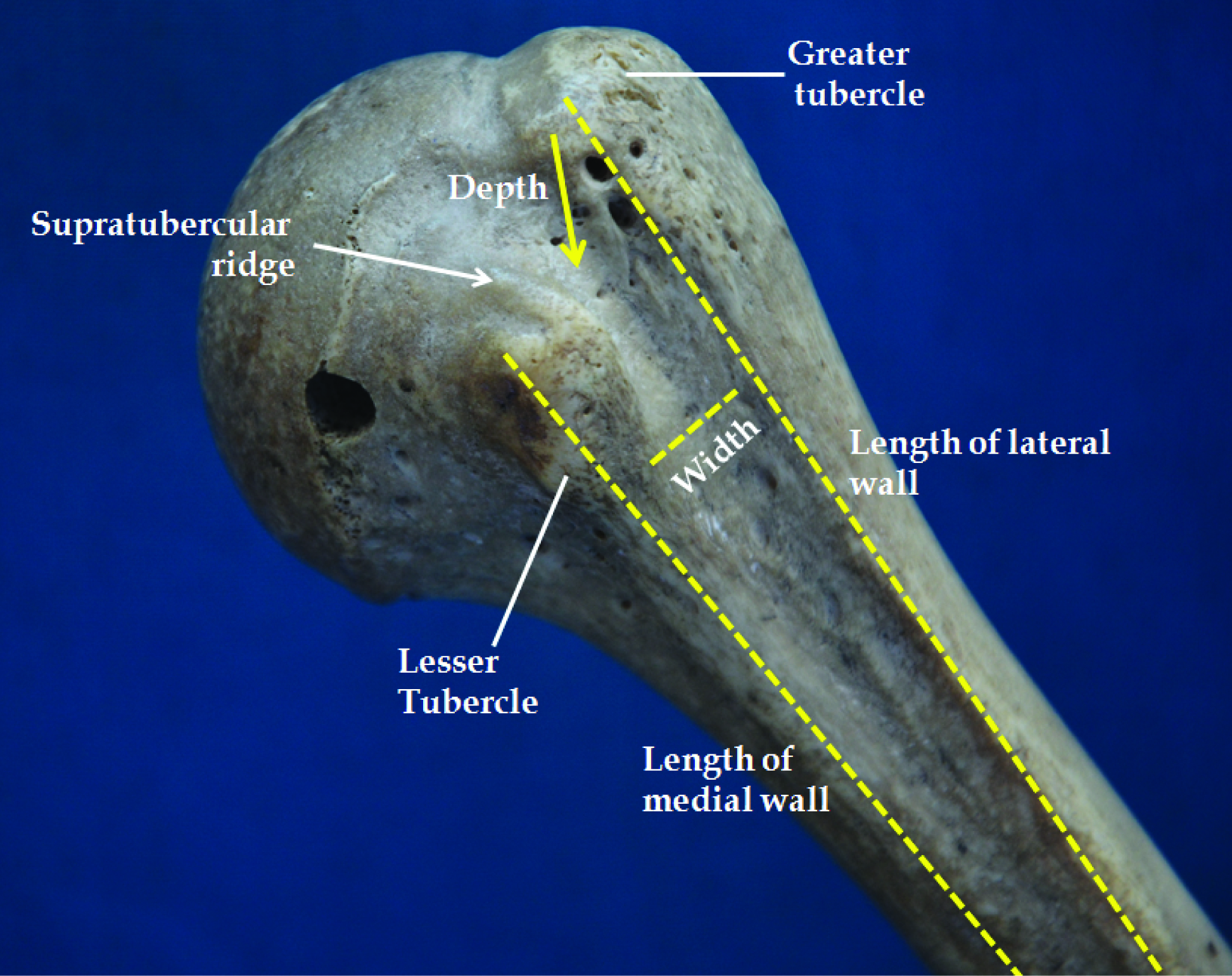
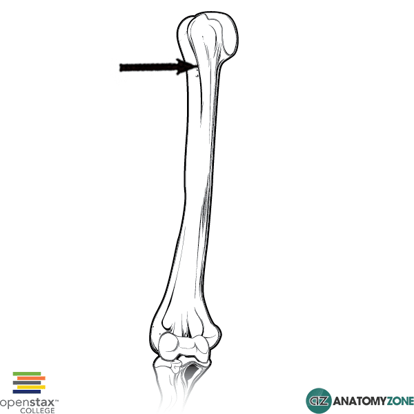
The bicipital groove intertubercular groove sulcus intertubercularis is a deep groove on the humerus that separates the greater tubercle from the lesser tubercle the bicipital groove lodges the long tendon of the biceps brachii between the tendon of the pectoralis major on the lateral lip and the tendon of the teres major on the medial lip.
Bicipital groove image. It was critical to distinguish between the cortical and endosteal surfaces during segmentation to ensure that the surface that comes in contact with the lbt was the one extracted fig. Biceps bicipital tendinitis is an inflammation of the long head of the biceps tendon as it passes through the bicipital groove of the anterior humerus see the image below. Images of the bicipital groove at 45 of external rotation were used in this study figure 2. It also transmits a branch of the anterior humeral.
The bicipital groove also known as the intertubercular sulcus or sulcus intertubercularis is the indentation between the greater and lesser tuberosities of the humerus that lodges the biceps tendon. The bicipital groove intertubercular groove sulcus intertubercularis is a deep groove on the humerus that separates the greater tubercle from the lesser tubercle the bicipital groove lodges the long tendon of the biceps brachii between the tendon of the pectoralis major on the lateral lip and the tendon of the teres major on the medial lip. It also transmits a branch of the anterior humeral. The longitudinal view was obtained with the probe resting perpendicularly to the bicipital groove.
To obtain a better image and eliminate the anisotropy effect the probe was adjusted to be parallel to the tendon for both the transverse and longitudinal views. The angular orientation of the bicipital groove has been referenced to the transepicondylar axis at about 55 in prior studies. 8 9 then standard line were drawn on the medial border of ulna from the tip of olecranon to the styloid process of the. Biceps tendinitis is a disorder of the tendon around the long head of the biceps muscle.
From these images the bicipital groove surface was segmented manually by a graduate student under the direction of a board certified radiologist.


