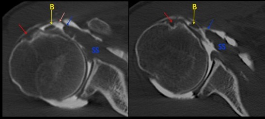
Bicipital Groove Ct

The tendon of the subscapularis muscle attaches both to the lesser tubercle aswell as to the greater tubercle giving support to the long head of the biceps in the bicipital groove.
Bicipital groove ct. Two views of a humeral head extracted from mri with the bicipital groove indicated. A type i normal tendon b type ii hourglass shaped hypertrophic tendon with extension of fraying into bicipital groove c type iii partial tear or fraying involving less than 50 of tendon width at the intraarticular region without fraying in the bicipital groove d type iv partial tear involving more than 50 of. Bicipital tendonitis is commonly diagnosed through clinical evaluation but confirmed through x ray mri or ct scan. The biceps tendon is contained in the rotator interval a triangular area between the subscapularis and supraspinatus tendons at the shoulder figure 1.
Bicipital groove x ray see more descriptions. Bicipital groove segmentation from ct data. This tool allows you to. Inflammation of the biceps tendon within the intertubercular bicipital groove is called primary biceps.
Bicipital groove segmentation from ct data. The lhbt can exhibit the magic angle phenomenon where it curves to enter the bicipital groove because it curves in the range of this artifact. Dislocation of the long head of the biceps will inevitably result in rupture of part of the subscapularis tendon. The bicipital groove is defined by the greater tuberosity lateral and the lesser tuberosity medial.
It serves to retain the long head of the biceps brachii. It serves to retain the long head of the biceps brachii. This produces intermediate signal intensity within the tendon that should not be mistaken for tendinosis or. Uk snomed ct clinical edition nhs data migration april 2020.
Biceps tendinitis is a disorder of the tendon around the long head of the biceps muscle. Classification of the long head of the biceps tendon by arthroscopic findings. Two views of a humeral head extracted from mri with the bicipital groove indicated. This was performed on 20 humeri ie 10 pairs to allow.
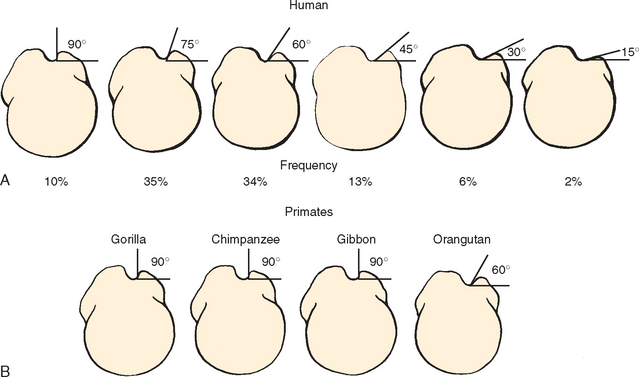
In this project we investigate the relationship between the 3d shape of the bicipital groove and the incidence of pathology of the long biceps tendon. The ct scan method and the computer assisted method enabled measurement of the distance between the central axis of the humeral head defined as the center of rotation of the humeral head articular surface and the posterior margin of the bicipital groove as performed by tillet et al. The bicipital groove is an osseous groove formed in the humeral head by the medial and lateral tuberosities.


