Bicipital Groove Anatomy Mri
Normal Mri Anatomy Of The Musculoskeletal System Radiology Key

A Profile View Of A Humeral Head Segmented From A Mri Of The

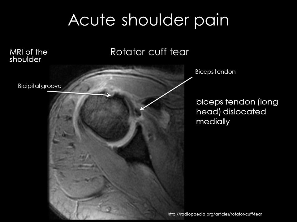
Long Head Of Biceps Tendon Dislocation Radiology Reference

Pathology Of The Long Head Of The Biceps Tendon Radsource
The Radiology Assistant Shoulder Anatomy Mri

The Radiology Assistant Shoulder Mr Anatomy
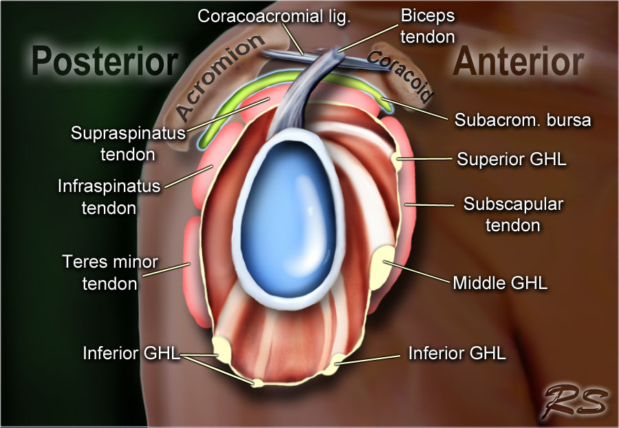
An accessory biceps tendon in the groove should not be mistaken for a tear.
Bicipital groove anatomy mri. It contains the tendon of the long head of the biceps brachii muscle which is ensheathed in a synovial reflection of the. In this project we investigate the relationship between the 3d shape of the bicipital groove and the incidence of pathology of the long biceps tendon. Potential causes of pulley lesions include a congenital rotator interval defect 23 a supratubercular ridge as an osseous protrusion of the lesser tuberosity 24 or a shallow groove 5. Bicipital tendinitis or biceps tendinitis is an inflammatory process of the long head of the biceps tendon and is a common cause of shoulder pain due to its position and function.
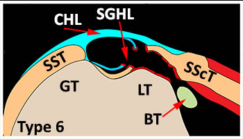
Bicipital groove green arrow in image below. Biceps tendon laxity in the bicipital groove may lead to superior extension of degenerative changes in the rotator interval 7 18 20. The accessory biceps tendon often originates from the supraspinatus tendon. The bicipital groove is typically 4 6 mm deep 1.
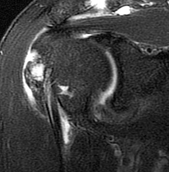
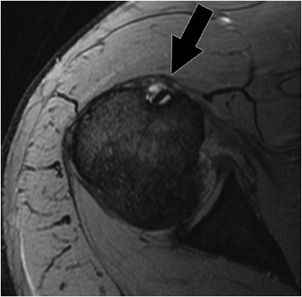
Image adapted from subscapularis footprint anatomy revisited with a 3 dimensional perspective and its relationship with the supraspinatus yoo j c et al. High t2 fluid signal intensity is noted around the thickened biceps tendon which is seen within its groove suggestive of tenosynovitis. Thickened with intermediate high t2 signal intensity noted at the supraspinatus tendon suggestive of tendinosis. Biceps tendinitis is a disorder of the tendon around the long head of the biceps muscle.
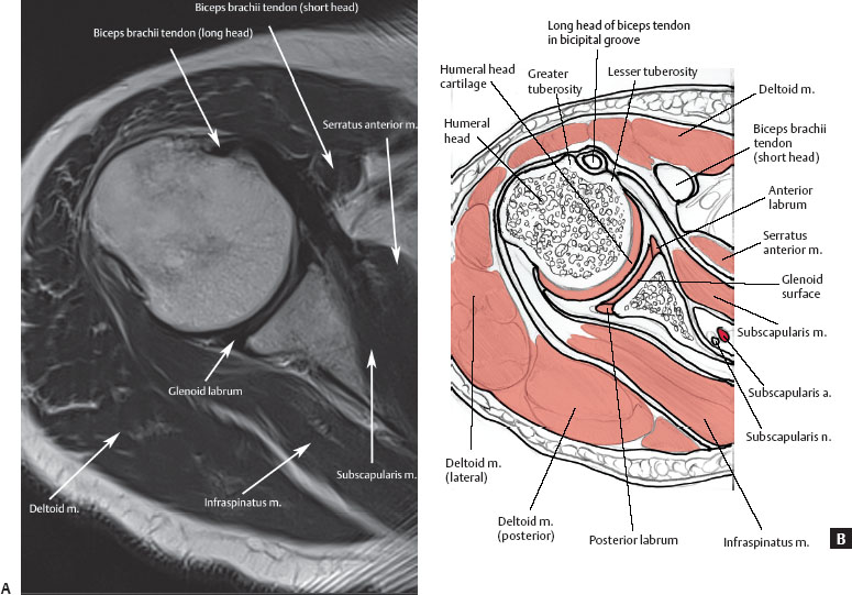
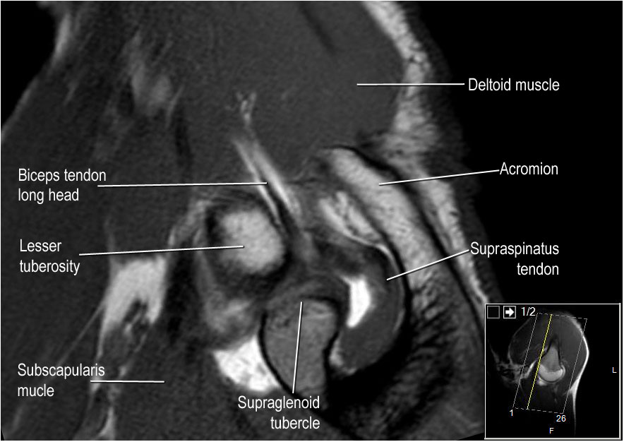
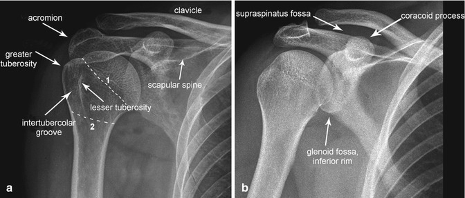
Part 1 normal anatomy and anatomic variants which begins on page 501. The bicipital groove intertubercular groove sulcus intertubercularis is a deep groove on the humerus that separates the greater tubercle from the lesser tubercle the bicipital groove lodges the long tendon of the biceps brachii between the tendon of the pectoralis major on the lateral lip and the tendon of the teres major on the medial lip. The bicipital groove is an osseous groove formed in the humeral head by the medial and lateral tuberosities. The important things to note from the anatomy we can use in imaging.
Both to the lesser tubercle aswell as to the greater tubercle giving support to the long head of the biceps in the bicipital groove. Shoulder anatomy mri normal anatomy variants and checklist robin smithuis and henk jan van der woude. It serves to retain the long head of the biceps brachii. The reader s attention is directed to part 1 accompanying this article titled pitfalls in shoulder mri.
Inflammation of the biceps tendon within the intertubercular bicipital groove is called primary biceps. It also transmits a branch of the anterior humeral. High t2 signal intensity noted at the of this humeral head at the biceps groove.
Pathology Of The Long Head Of The Biceps Tendon Radsource

Long Head Of Biceps The Biomechanics Of Injury Functional
Https Www Advancedbodyimaging Org Portals 9 Meetings 2010 Summer Steinbach Mri 20of 20the 20shoulder 20 20long 20head 20of 20the 20biceps 20tendon Pdf

Figure 5 From Mri Of The Rotator Interval Of The Shoulder

Pathology Of The Long Head Of The Biceps Tendon Radsource
The Radiology Assistant Shoulder Anatomy Mri
Long Head Of The Biceps Tear A Mri Arrow Bicipital Groove With
Pathology Of The Long Head Of The Biceps Tendon Radsource
Msk Clinical Cases Traumatology Ppt Video Online Download

Artifacts And Pitfalls In Shoulder Magnetic Resonance Imaging
Joints Radiology Key
Shoulder Mri Radiology Key

A Profile View Of A Humeral Head Segmented From A Mri Of The
